How does the brain impose boundaries on free recollection?

Try to list the names of Hollywood stars. Julia Roberts and Richard Gere might spring to mind. But other names – say Albert Einstein or the name of your first-grade teacher – won’t come up. We do this so easily that our ability to recall information that fits into a particular category seems trivial. But until now, the brain mechanism that allows us to confine memory retrieval to a particular class or category was not known. New research in the lab of Prof. Rafael Malach of the Neurobiology Department of the Weizmann Institute of Science, in collaboration with the lab of Prof. Ashesh D. Mehta of The Feinstein Institute for Medical Research, which was recently published in Nature Communications, has provided direct insight into the activity of nerve cells in the human brain’s cortex as memories are retrieved in this bounded manner.
Now imagine yourself on a crowded beach in summer, looking for a lost friend among all the sunbathers and picnickers. Even with all of the distraction around, you pick him out from a fair distance by the red shirt he was wearing. How did you manage to focus on this one detail among all those in your environment? When we are looking for something specific, we have an “attention mechanism” that “warms up” the visual center in the brain that represents this object. That is, those particular neurons become more active, so the second the person or thing we are looking for pops into view, our attention can immediately be focused on its presence.
Portraits only, please
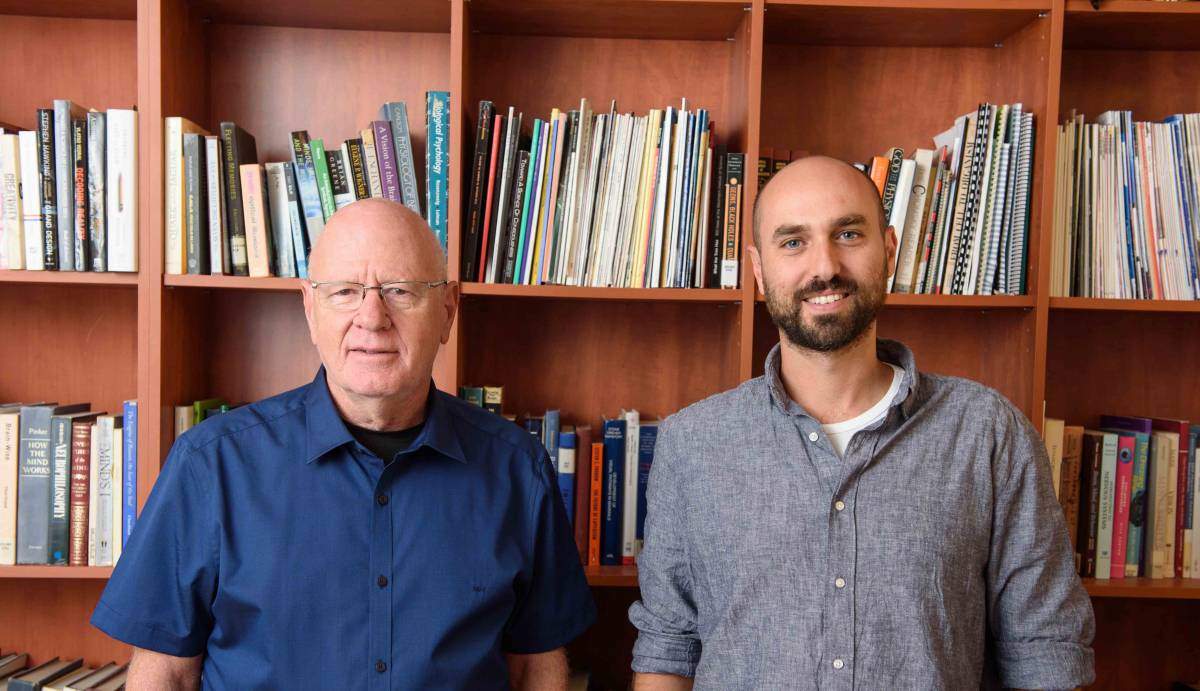
Yitzhak Norman, the research student in Malach’s lab who led the study, asked whether trying to find information stored in our memory is similar to the process by which we identify objects we are looking for in our surroundings. This hypothesis, which is not new, is called “attention to memory,” or AtoM for short. However, until now, scientists have not had a good way to observe this type of internal memory search in real time in the human brain, or to unlock the mechanisms governing this process.
The present study, explains Norman, made use of the basic organization of the human visual cortex: “There is an anatomical division in the brain’s visual system. Faces, body parts, places and objects: each are represented in different sub-regions of the visual system. This spatial separation can help us measure the selective recall of these various objects.” To measure this activity in the brain, the researchers turned to epilepsy patients. These patients – who all volunteered for the study – have a series of electrodes implanted in their brains for a period of about two weeks as an integral part of their clinical epilepsy diagnosis. While they are in the clinic waiting for an epileptic attack to occur, these patients often are willing to participate in cognitive experiments that give researchers precise data about neuronal activity in the brain that cannot be attained by any other method.
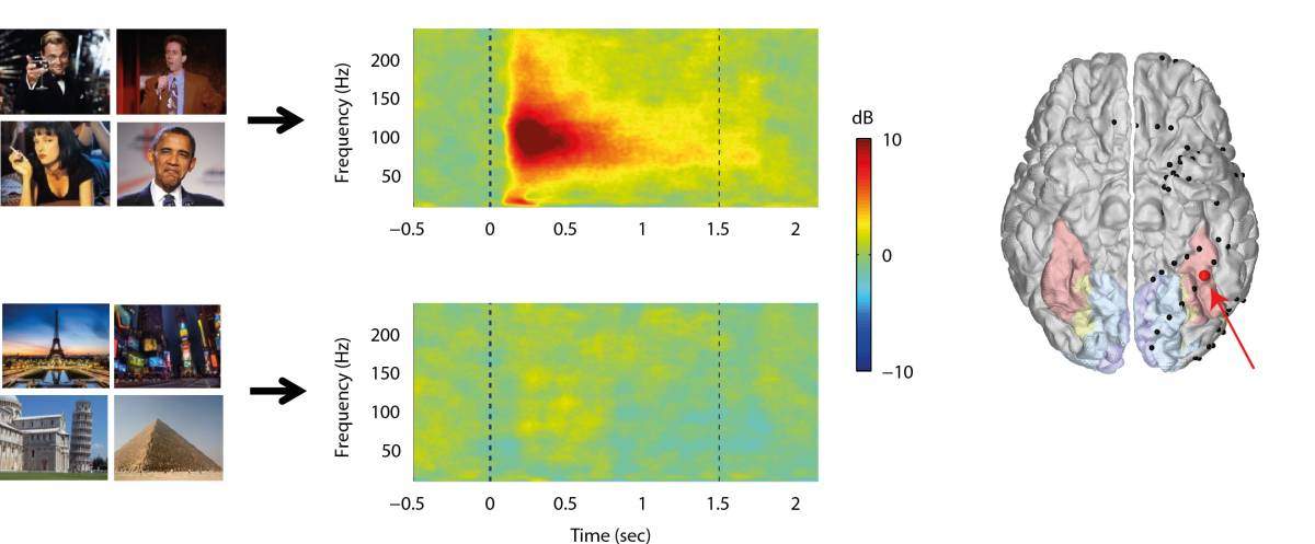
The activity in the visual area specializing in faces “warmed up” in the same way it does when we search for an object in the external world
In the half-hour experiments, the patients were shown a series of photos, in random order, including of famous individuals (Barack Obama, Uma Thurman, Bart Simpson, etc.) and famous monuments (the Tower of Pisa, Statue of Liberty, Golden Gate Bridge, etc.), and asked to remember them in as much detail as possible. A few minutes later, they were asked to recall the photos they had seen – but they were asked to think of each category separately: only portraits in one session, and only landscapes in the other. The recordings of their brains’ activities revealed that when the patients attempted to recall the portraits, the activity in the visual area specializing in faces “warmed up,” in the same way it does when we search for an object in the external world. The other regions of the visual cortex showed no change in activity, so this warming up was, indeed, selective. When asked to recall the landmark photos, the activity in the area for faces was quiescent, while the part in the brain specializing in images of places warmed up. This mechanism is fast – raising the level of neuronal activity within the first second that an area is called upon – and stable, with the enhanced activity remaining constant throughout the recall session.

The findings might be significant for treating people suffering from memory loss after injury. Norman: “If we understand the mechanism, we might be able to develop alternative cognitive strategies for improving recall, or even micro-stimulation techniques to therapeutically improve retrieval.” According to Malach, the findings may also help to highlight the more general mechanism that enables us to control cognitive processes that occur spontaneously. This, in turn, may help us to shed new light on the fundamental tension that exists between the spontaneous nature of our thinking and the process that enables us to reign in this potentially chaotic activity to its optimal boundaries.
Prof. Rafael Malach’s research is supported by the Murray H. and Meyer Grodetsky Center for Research of Higher Brain Functions, which he heads; the Lulu P.and David J. Levidow Fund for Alzheimers Diseases and Neuroscience Research; the Dr. Lou Siminovitch Laboratory for Research in Neurobiology; the Leff Family; the Appleton Family Trust; and the estate of Florence and Charles Cuevas. Prof. Malach is the recipient of the Helen and Martin Kimmel Award for Innovative Investigation; and he is the incumbent of the Barbara and Morris L. Levinson Professorial Chair in Brain Research.

Recent Comments