A new MRI method developed at the Weizmann Institute of Science improves our ability to study the brain and other non-homogeneous tissues
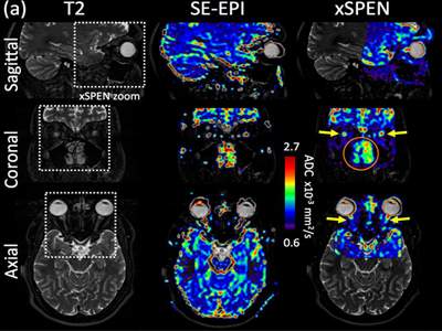
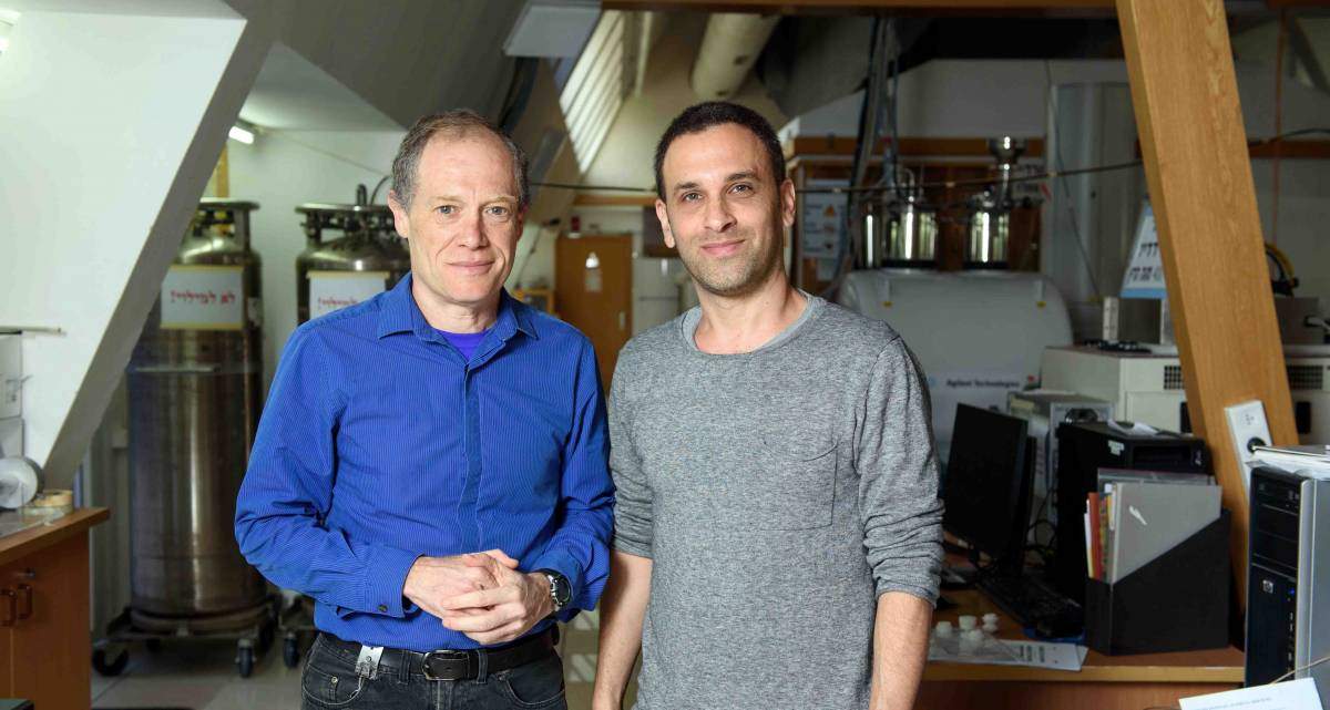
Magnetic resonance research at the Weizmann Institute of Science has been at the forefront of the field for decades. Four new magnetic resonance scanners, all characterized by the strongest magnetic fields commercially available, are in various stage of installation and start-up at the Institute. These could open unprecedented opportunities for biological research and clinical translation. “But some tissues and organs still present considerable challenges when we attempt to tackle them with MRI at these new, higher fields. We are very active in developing methods to overcome those challenges,” says Prof. Lucio Frydman of the Institute’s Chemical and Biological Physics Department.
Frydman explains that MRI works best on tissues that are homogeneous – meaning that water molecules and metabolites reside in uniform regions within the magnetic field that creates the MR images. But not all regions in the body are homogeneous: inhomogeneity appears, for example, at the interfaces between nasal passages, sinuses and the brain; close to the eyes, in the basal region of the brain, close to the optic nerves and the olfactory bulb; and throughout most of the abdomen. The MRI magnets generally fail to compensate for the field distortions arising from these heterogeneities, and this gets exacerbated in proportion to the strength of the applied field, threatening to rob some of the new ultrahigh field scanners’ potential. Such handicaps become particularly noticeable in MRI techniques that rely not just on localizing molecules but on mapping their diffusion – that is, in mapping the extent and direction of water movements in tissues. Such diffusion analyses can highlight small tumors or neuronal development; and in non-homogeneous tissues at very high fields. However, these measurements usually present corrupted information.
Embracing the limitations
Frydman and his team have worked for nearly a decade developing new approaches to tackling these problems, including a so-called xSPEN (for cross-term SPatiotemporal ENcoding) experiment that overcomes inhomogeneity issues, producing MRI diffusion images in a very robust way. To create xSPEN, the team manipulates all the critical stages of an MRI scan: There is a new pre-encoding of the information before the scan begins; the scan itself, including customized shapes of the field gradients that elicit the images from the water; and a new data-processing algorithm that had to be developed to convert the MRI signals into images. “There was quite a bit of programming involved, but developing these new techniques required first and foremost breakthroughs in the physical understanding of alternative ways of performing MRI,” says Frydman. “The result is a new approach to MRI that can deliver more accurate images because, for the first time, it deals with inhomogeneities by including them in the image-generating process, rather than by trying to forcibly overcome them or compensate for them with post-acquisition image corrections.”
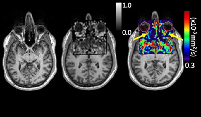
A demonstration of the method, conducted on a preclinical system and then on healthy human volunteers in a clinical scanner, was recently published in Scientific Reports. Among other features, the scans clearly show xSPEN’s ability to target challenging regions in the periphery of the brain and throughout the human head, including the thin optic nerve leading from the eye into the brain. These studies and technologies are now being extended in Frydman’s group to investigate other regions and organs − including the fetal-placental unit − with the aim of understanding how nutrients are shared or transferred between mother and fetus.
X-SPENding effort in the clinic
In parallel with these, Frydman and his team have begun working with two hospitals in Israel in order to apply their methods to scanning challenging regions in patients. Their objective is to combine all the steps in their new data-acquisition and processing protocol into one easy-to-use package that can be installed in a hospital MRI facility and operated by technicians with minimal extra training.
“The great thing is that despite their new way of delivering MR images, these scans are entirely compatible with existing hardware,” says Frydman. “They are also non-invasive and can be merged into existing clinical protocols and optimized under realistic scanning conditions.”
Future challenges and opportunities
The Institute’s MR activities – electron paramagnetic resonance (EPR), nuclear magnetic resonance (NMR) and magnetic resonance imaging (MRI) – are in the midst of singular upgrades. These include the installation of a state-of-the-art 15.2 Tesla (T) MRI scanner for animal research, the addition of brand new 3 T and 7 T scanners for use in humans, and a 23.5 T (1 GHz) machine for NMR research.
Frydman is highly optimistic regarding the future of MRI: “In the latest trials, for instance, we observed that the new 15.2-T system provides a ten-fold sensitivity improvement over what we had previously recorded in our lab using a state-of-the-art 7-T system. This is a tremendous advantage, and now it’s time to leverage it without compromising on any other aspect that MRI has already achieved.”
Various MRI groups at the Weizmann Institute plan to use these new machines for a range of additional projects – from developing new biosensors for the early detection of disease, to tracking of how new vasculatures are established and shedding light on the workings of the brain. Technological and scientific MRI applications are happening hand-in-hand at the Weizmann Institute of Science and, as these synergies develop and grow, there are bound to be new and exciting biological breakthroughs and clinical improvements.
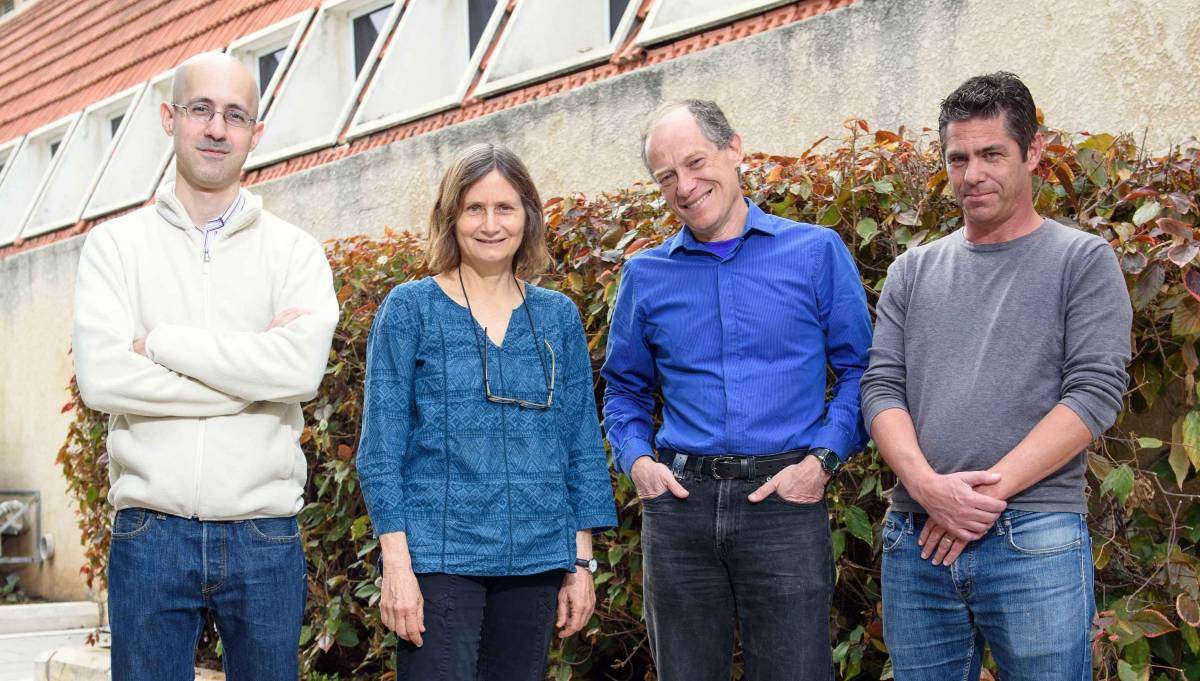
Prof. Lucio Frydman’s research is supported by the Helen and Martin Kimmel Institute for Magnetic Resonance Research, which he heads; the Clore Institute for High-Field Magnetic Resonance Imaging and Spectroscopy, which he heads; the Helen and Martin Kimmel Award for Innovative Investigation; the Leona M. and Harry B. Helmsley Charitable Trust; the Adelis Foundation; the Dr. Dvora and Haim Teitelbaum Endowment Fund; the Comisaroff Family Trust; the Emil and Rita Weissfeld Family Foundation; the Takiff Family Foundation; and the European Research Council. Prof. Frydman is the incumbent of the Bertha and Isadore Gudelsky Professorial Chair.

Recent Comments